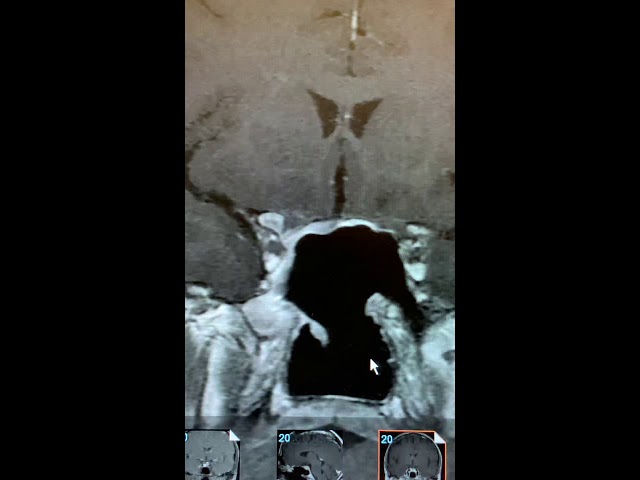
This is the fourth in a series of pituitary MRI’s. This video describes the normal sinonasal anatomy and provides a stunning description of how this area is affected after endoscopic pituitary surgery. The comparison and visuals, coupled with Dr. Blevins explanations, are quite compelling.
Stay tuned for more videos in this series coming later this week.
Click here to watch the other MRI videos in the series.
© 2019 – 2022, Pituitary World News. All rights reserved.